

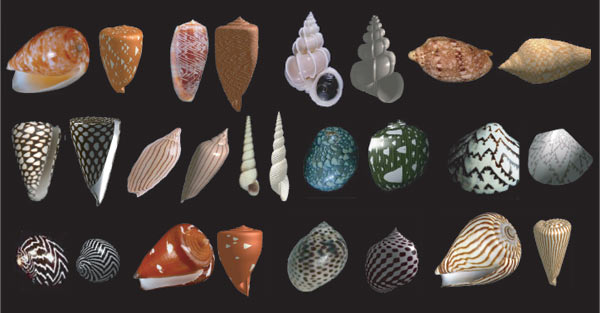
 By adjusting nine parameters in a single equation, a computer model can generate patterned shells (right example in each pair above) that closely resemble real mollusk shells. (Alistair Boettiger/UC Berkeley)
Sea mollusks taste their memories to build shells
| 01 April 2009
BERKELEY — University of California, Berkeley, graduate student Alistair Boettiger has amassed a beautiful collection of seashells, but not by combing the beach. He created them in his computer.
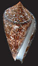 A simple neural network model of seashell growth can generate realistic mollusk shells based on a simple principle discovered 140 years ago. The computer-generated shell at left closely resembles the real shell of the mollusk Conus gloriamaris (above), a poisonous marine snail found throughout the Indian and Pacific oceans. (Alistair Boettiger & George Oster/UC Berkeley; Bard Emmentrout/Pitt) |
The "neural net" model explains how mollusks build their seashells based on the finding that the mollusk's tongue-like mantle, which overlaps the edge of the growing shell, senses or "tastes" the calcium carbonate layer laid down the day before in order to generate a new layer.
"The pattern on a seashell is the mollusk's memories," said Oster, a professor of environmental science, policy and management and of molecular and cell biology. "The shell is laid down in layers, so the mantle is sensing the history of the mollusk's 'thoughts' and extrapolating to the next layer, just like our brains project into the future."
The studies may help neuroscientists understand how neural networks work in the brain and throughout the body, where neural nets cover our skin and all internal organs.
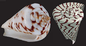
The researchers' computer model can reproduce a wide variety of shell shapes, colors and patterns, including Conus vicweei (above) and Olivia porphyria. In both photos, the real shell is at left and the computer-generated image is at right. (Alistair Boettiger/UC Berkeley)
"The model gives us a remarkable ability to explain much of the dramatic diversity of both shape and pattern that we see in the natural world," Boettiger said.
To build their model, the UC Berkeley scientists first studied electron microscope images of mollusk mantles in order to understand the network of neurons connecting the sensing cells in the mantle with the secretory cells that produce calcium carbonate and proteins - many of them colored pigments - incorporated into the growing shell. Different rates of calcium carbonate secretion determine the shape of the spiral, while different amounts of pigment secretion create a pattern unique to each species.
They then modeled the size of the excitatory and inhibitory regions surrounding the secretory cells and the cells' firing thresholds - nine parameters in all - as a neural network that determines how much calcium and pigment is secreted.
Based solely on these nine parameters, Boettiger, Oster and Ermentrout were able to reproduce the shapes and patterns of almost every known sea mollusk.
Interestingly, they found that all shell patterns fall into three basic classes: stripes perpendicular to the growing edge, bands parallel to the growing edge, and complex patterns created by asymmetric "traveling waves" of pigment or calcium deposition.
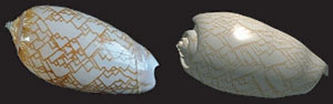
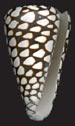 The computer-generated shell at left closely resembles the shell of the marbled cone (above), a poisonous snail found in Hawaii and throughout the Indo-Pacific. (Alistair Boettiger & George Oster/UC Berkeley; Bard Emmentrout/Pitt) |
Famed computer scientist Alan Turing showed in 1952 how local activation/lateral inhibition could work chemically, and biologist Hans Meinhardt used this chemical model to create realistic seashell patterns in the 1970s, which he published in a 1995 book called "The algorithmic beauty of sea shells."
At that time, the neural basis of shell patterning hadn't been widely accepted, though Oster and Ermentrout published an earlier version of the neural model in the 1970s. One problem with Meinhardt's chemical model, which hypothesized reactions among chemicals diffusing through the snail shell, is that it required different chemical reactions to produce each shell pattern.
"Our real contribution is not reproducing the patterns, but showing that the nervous system can do it with one equation based on the principle discovered by Ernst Mach in the 1860s," Oster said.
Striped shells are the easiest to explain with this neural network model. A pigment-secreting cell inhibits secretion of pigment by neighboring cells but not itself, so that the same pattern is repeated day after day, yielding a stripe. Similarly, if one cell pumps up calcium carbonate secretion while depressing secretion by surrounding cells, ridges result. Interestingly, the stripes or ridges split naturally as the shell grows, a mathematical necessity because the size of the inhibition area remains the same as the shell's edge grows.
Bands parallel to the growing edge can be explained by inhibition of future activity. Pigment secreted on one day can inhibit secreting cells for a few days, resulting in an on/off pattern that produces a series of bands.
The most interesting patterns, however, are waves of activity that interfere to produce zigzags, diamonds, chevrons, arrowheads and a host of other shapes. These come about when a pigment inhibits future secretion at that site but excites secretion in surrounding cells. The pigment thus moves laterally on successive days, producing the equivalent of a traveling wave.
Ironically, most sea snails don't care a whit about their shell pattern. They are buried in the mud of the seafloor where their patterns are hidden even from potential mates.
"The pigment is a cue to get the mantle in register so it builds the right shaped shell, and is only an epiphenomenon reflecting neural activity," Oster said. "It is incidental to the snail."
"There is no strong selective pressure to drive patterns, so evolution can explore the entire parameter space" of possible shells, Boettiger added. "That was one rewarding thing about this work; it brought some nice aesthetics to the whole project."
With their success describing shell patterning, Oster plans to move on to his real interest, how cuttlefish rapidly change their patterns in response to the environment. Cuttlefish see a pattern in the environment and alter their skin pattern to blend in, he said, often flickering so rapidly that they resemble an hypnotic strobe.
The work was supported by the National Science Foundation.